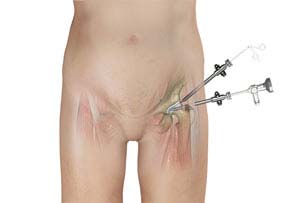
Hip Arthroscopy

Hip arthroscopy, also referred to as keyhole surgery or minimally invasive surgery, is performed through very small incisions to evaluate and treat a variety of hip conditions. Arthroscopy is a surgical procedure in which an arthroscope is inserted into a joint. Arthroscopy is a term that comes from two Greek words, arthro-, meaning joint, and -skopein, meaning to examine. Arthroscope is a pencil-sized instrument that has a small lens and lighting system at its one end. Arthroscope magnifies and illuminates the structures inside the body with the light that is transmitted through fiber optics. It is attached to a television camera and the internal structures are seen on the television monitor.
Hip arthroscopy may be indicated in following conditions:
- Debridement of loose bodies: Bone chips or torn cartilage debris cause hip pain and decreased range of motion and can be removed with hip arthroscopy.
- Removal of adhesions: Adhesions are areas of built up scar tissue that can limit movement and cause pain.
- Repair of torn labrum: The labrum lines the outer edge of the “socket” or acetabulum to ensure a good fit. Tears can occur in the labrum causing hip pain.
- Removal of bone spurs: Extra bone growth caused by injury or arthritis that damages the ends of the bones cause pain and limited joint mobility.
- Partial Synovectomy: Removal of portions of the inflamed synovium (joint lining) in patients with inflammatory arthritis can help to decrease the patient’s pain. However, a complete synovectomy requires an open, larger hip incision.
- Debridement of joint surfaces: Conditions such as arthritis can cause the breakdown of tissue or bone in the joint.
- Repair after Trauma: Repair of fractures or torn ligaments caused by trauma.
- Evaluation and diagnosis: Patients with unexplained pain, swelling, stiffness and instability in the hip that is unresponsive to conservative treatment may undergo hip arthroscopy for evaluation and diagnosis of their condition.


